Research within WITNe is conducted across labs that communicate openly. Our engineering researchers, medical researchers and medical practitioners work closely together, sharing knowledge, resources, people, collaborator networks, animal models, grant support, access to industry contacts, training opportunities and increased visibility.
WILLIAMS LAB

Justin Williams
Professor, BME

MISSION
Develop new devices for recording from and stimulating neural tissue that are both safe and durable for long-term implantation, as well as safe for use in humans and animals; plus, validate the use of these new devices in animal models and human patients.
WHAT WE’RE UP TO
- Developing new devices for recording from and stimulating neural tissue
- Designing these devices to be both durable for long-term implantation and safe for use in humans and animals
- Using these technologies in a variety of situations, from use in a basic physiology lab recording from single neurons, to clinical settings where people with motor disabilities might benefit from a brain-computer interface or other neural prosthetic communication device
LUDWIG LAB
Kip Ludwig
Professor, School of Medicine and Public Health

MISSION
Translate next generation neuromodulation therapies to highjack the nervous system to treat circuit dysfunction and deliver biomolecules to target areas with unprecedented precision.
WHAT WE’RE UP TO
- Acute and longitudinal benchtop characterization of new minimally invasive neural interfaces based on proven experience supporting FDA regulatory submissions
- The application of cutting-edge optical tools in rodents to better understand and design clinical neuromodulation interfaces
- Comprehensively instrumented swine model to refine new neuromodulation strategies at human anatomical scale in order to assess effects and side effects
- Detailed functional and post-mortem anatomical evaluation across animal and human experimental models to reduce the time to translation of new concepts


SUMINSKI/LAKE LAB

Aaron Suminski
Senior Scientist, Department of Neurological Surgery

Wendell Lake
Assistant Professor, Department of Neurological Surgery


MISSION
Understand how neuromodulation therapies regulate neural activity both in close proximity to the stimulating electrodes and in neural circuits receiving projections from the site of stimulation.
WHAT WE’RE UP TO
- Optimizing patient outcomes following implantation of neuromodulation therapies using advance imaging techniques and electrophysiology
- Investigating the mechanisms of action for neuromodulation therapies using experimental models across the translational spectrum from basic science in rodent models to clinically translatable research in large animal models and human patients
HAI LAB
Aviad Hai
Assistant Professor, Department of Biomedical Engineering
Fellow, Grainger Institute for Engineering

MISSION
Engineer minimally invasive tools to access the nervous systems for neurobiological studies of brain function.
WHAT WE’RE UP TO
- Developing nanoscale electrochemical and electromagnetic sensors for recording neurophysiological activity and neurotransmitter in the brain
- Combining them with neuroimaging modalities such as functional Magnetic Resonance Imaging
- Using microelectronic fabrication, nanolithography, and surface chemistry techniques, together with in vitro and in vivo animal models to establish our technologies for clinical applications in neurology


LOCI LAB

Kevin Eliceiri
Director and Principal Investigator, Laboratory for Optical and Computational Instrumentation (LOCI) Laboratory
Associate Professor, Department of Medical Physics and Department of Biomedical Engineering

MISSION
Develop advanced optics and computational techniques for imaging and experimentally manipulating living specimens
WHAT WE’RE UP TO
- Computational optics research
- Imaging the cellular microenvironment of wound healing models
- Developing software tools such as FIIJI for 3D visualization analysis
BHASKAR LAB

Dhananjay Bhaskar
Assistant Professor, BME
MISSION
We are a computational lab with interests in machine learning, mechanistic modeling, and AI for Science. Our goal is to understand how structure and dynamics give rise to function in complex systems – from molecules and cells to circuits and behavior.
We develop methods that capture the shape, symmetry, and flow of biological processes in high-dimensional, often noisy data. To do this, we design algorithms in geometric deep learning, topological data analysis, and manifold learning, that relate the ‘shape’ of data to its underlying biology. We tackle challenging inverse problems – using data to infer the cellular and molecular interactions or neural connectivity that drive observable behavior. Ultimately, our aim is to create biologically informed, interpretable, AI models that not only explain how systems work but also drive translational impact through engineering and precision medicine.
WHAT WE’RE UP TO
- Identifying in-silico biomarkers of neurological and psychiatric conditions using multimodal neural and behavioral data
- Integrating and translating across brain imaging modalities
- Learning high-order interactions and network dynamics from large-scale brain activity recordings
- Building brain encoding and decoding models to understand how information is represented, transformed, and recalled
- Designing new learning algorithms that emulate neuronal dynamics, plasticity, and self-organization for more biologically grounded AI
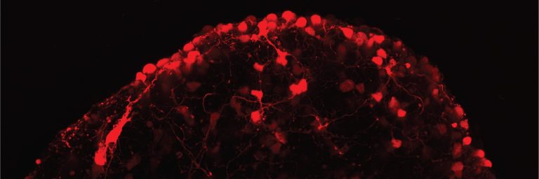
NERV LAB
Jonathan Soucy
Assistant Professor, BME
MISSION
We engineer neural tissues and interfaces to restore vision in degenerative retinal diseases.
The Neural Engineering for Restoring Vision (NERV) lab studies how the visual system develops, remodels, and can be repaired after injury or disease. We engineer ex vivo and stem cell-derived models of the retina, optic nerve, and retinorecipient targets in the brain to uncover the principles of neuroplasticity. By combining electrophysiology, tissue engineering, and cell transplantation, we are developing tools to study how transplanted neurons integrate into host circuits and using transplantation itself as a strategy to restore vision. Our goal is to translate fundamental discoveries into therapies that promote remodeling, repair, and recovery in the visual system.
Visit the NERV lab website: nervlab.bio
WHAT WE’RE UP TO
- Organoid and organ-chip models – Engineering retinal tissues to study neural circuit formation and donor-host integration
- Neuron transplantation therapies – Developing strategies to restore vision lost to glaucoma and other optic neuropathies
- Activity modulation tools – Using chemogenetic and optogenetic approaches to tune donor and host neuron activity and study neuroplasticity
- High-density electrophysiology – Applying advanced recordings to map connectivity and assess the recovery of vision after transplantation